|
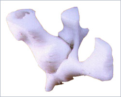
This is a case of
a 24 year old man who had met with a vehicular accident
and fractured his hip. The hip is a ball (femoral head)
and socket (acetabulum) type joint and in this case,
the acetabulum was fractured into 4 pieces. The reduction
of this fracture was difficult because it was located
on the posterior side and also one of the smallest fractured
piece had been grossly dislocated and hence was obstructing
the normal process.
The 3D CT scan did show this
small piece but it could not delineate the origin of
this piece. The model enabled to locate the origin of
this piece. A few pre-operative trials were done on
the model to arrive at its proper orientation. This
reduced the surgery time and possibly avoided additional
surgeries as well.
|


